
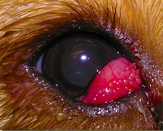
Cherry Eye in Dogs, medically known as the prolapse or eversion of the gland of the Nictitating Membrane (Third Eyelid), is a condition in which the gland of the lower eyelid bulges out and appears as a bright reddish mass in the lower corner of the eyes. Just because of this reddish mass bulging out from the eye, the disease is also known as “Cherry Eye Disease”.
The exact cause and causative agent of Cherry Eye Disease is still known. But it’s hereditary and genetics probably play a large role. While cherry eye can affect one or both eyes, mostly it doesn’t happen in both at the same time. Affected dogs are uncomfortable from eye dryness, swelling, irritation, inflammation and pain. They rub their faces on flooring or furniture to try and relieve discomfort. But this disease can be treated easily and prognosis is always positive.
Anatomically, each eye of domestic dogs contains a nictitating membrane – also known as a “third eyelid” – which hides beneath the lower eyelid and normally is not visible. Tear glands are located around the cartilage connections of the nictitating membranes and provide a major source of tear film and eye lubrication. However, if the fibrous tissues that hold the third eyelids to the globes of the eyes sometime become weakened, the tear glands can bulge out (“prolapse”) over or around the third eyelid, appearing as dangerous-looking bright red masses.
How Cherry eyes Disease Affects the Dogs:
Cherry eye can occur in just one of a dog’s eyes (unilaterally) or sometimes in both eyes (bilaterally). Dogs that develop cherry eye usually show symptoms of ocular irritation, dryness, redness (conjunctivitis), swelling, inflammation and pain. The vision of dogs with cherry eye may be adversely affected as well, especially if the surface of the affected eyes becomes scratched, infected or abraded with rough surface rubbing.
Symptoms of Cherry Eye in Dogs:
Cherry Eye Disease don not develop or start progressively. It appear like a sudden abnormality. And owner will observe a continuously protruding red mass in the lower corner of eye all of a sudden. The most obvious sign of cherry eye is a well-defined solitary mass of red tissue bulging from the inner corner of one or both of a dog’s eyes. Often, this protrusion is the only observable sign that owners see.
If cherry eye is not treated and corrected within a reasonable period of time, the dog can develop additional and sometimes rather serious ocular complications. When the third eyelid and tear gland are not in the proper place, the eye can become red, dry, irritated and inflamed. There may be abnormal watery discharge from affected eyes. Some dogs rub their eyes with different surface and they may aggravate the severity of disease. Owners of dogs with cherry eye may notice one or more of the following:
- Eye redness (conjunctivitis)
- Swelling around the eyes
- Tear production – signs of eye drainage
- Abnormally dry eyes – insufficient tear production
- Rubbing/pawing at the eyes
- Squinting
- Vision impairment
- Other signs of eye irritation.
Dogs/Breeds Commonly Affected by Cherry Eye Disease:
It is more prevalent in young dogs and dogs with age of 6 months to 2 years are mostly seen affected. Some breeds develop this condition more commonly than other dogs. These breed are
- Cocker Spaniels
- Bulldogs, Beagles
- Bloodhounds
- Lhasa Apsos
- Shih-Tzus
- Brachycephalic Breeds (dogs with very short flat faces and wide heads)
Diagnosis of Cherry Eye Disease:
Cheery Eye diagnosis is not so difficult for a veterinarian. Your veterinarian will detect in the very first look at bulging red mass. Although some veterinarian suggest for some biopsy examinations in old dogs because they may develop this condition due to some carcinomas.
Treatment Cherry Eye in Dogs:
Cherry Eye should be treated as soon as possible. Because the condition itself is not particularly so dangerous to dogs but delay in treatment may lead to more serious secondary problems. The longer the that the third eyelid gland is out of place and exposed to environmental elements, the more inflamed, irritated and possibly infected it may become. The main objective for the treatment of cherry eye disease are:
- Return the function and appearance of the third eyelid structures to as normal a state as possible
- Reduce abnormal discharge from the affected eye(s)
- Minimize irritation and injury to the corneal and conjunctival tissues
- Preservation of tear gland of the third eyelid
- Minimize the risk of secondary bacterial infections
- Minimize the dog’s discomfort via medication or surgery
Treatment Options for Cherry Eye Disease:
Two type of option are available for the surgical treatment of Cherry eye Disease.
- Removal of the protruding gland of third eyelid
- Re-positioning of the tear gland to its normal position
Re-positioning of the gland to its normal position is the best choice. Veterinarian use different surgical techniques and the bulging part is sutured back to its normal position. This approach save the tear gland and the eyes don’t become dry.

Removal of the gland may cause more problems e.g. drying of the eye and that further need medicinal treatment.
Prevention of Cherry Eye in Dogs:
The precise cause of cherry eye is unknown. However, because it appears more often in certain breeds, it is thought to have a genetic component involving weak connective tissue around the third eyelid. Inflammation and hypertrophy seem to play a role as well.


